
Spinal Anatomy Spinal Regions Bones and Discs Vertebrae Spinal Cord
Human back. The human back, also called the dorsum ( pl.: dorsa ), is the large posterior area of the human body, rising from the top of the buttocks to the back of the neck. [1] It is the surface of the body opposite from the chest and the abdomen. The vertebral column runs the length of the back and creates a central area of recession.

Diagram Of Backbone The Vertebral Column Anatomy And Physiology I Once the topic is
What does the spine do? Your spine has several important functions, including: Giving your body structure (shape). Supporting your body (posture). Protecting your spinal cord (nerves that connect your brain to the rest of your body). Allowing you to be flexible and move. Anatomy Where is the spine located?
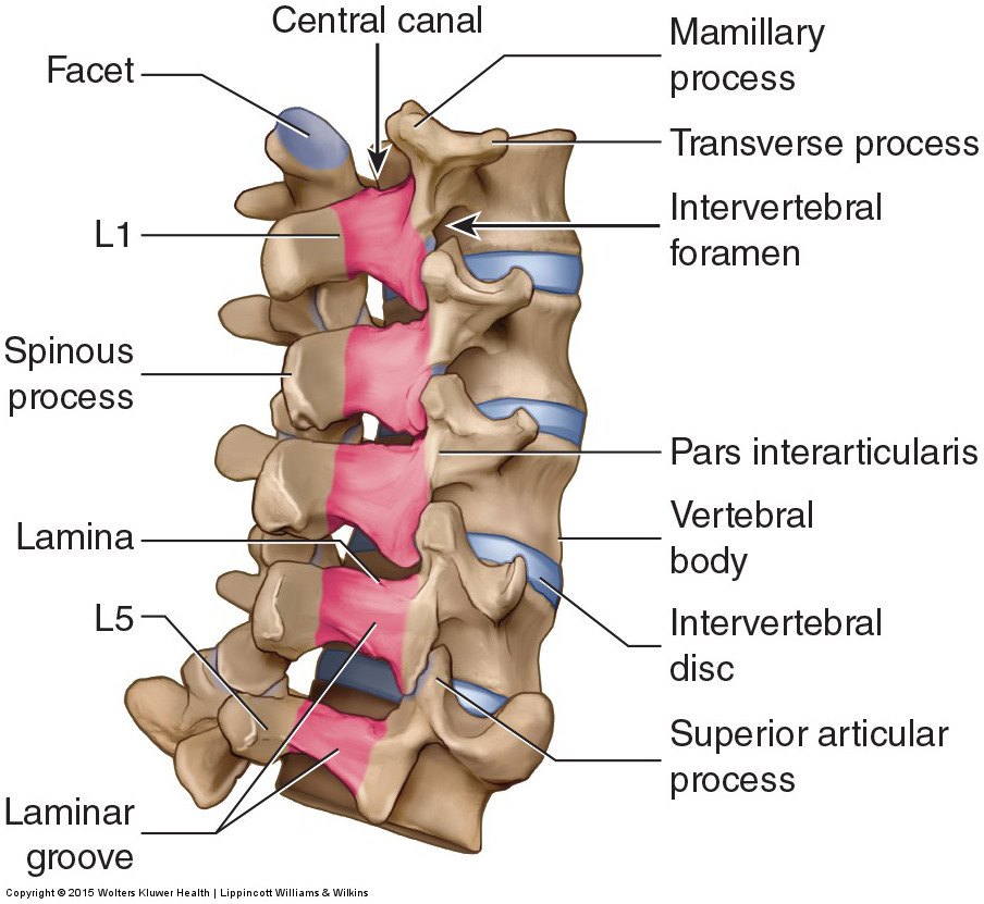
Diagram Of Human Backbone Anatomy Of Spine And Neck Anatomy Drawing Diagram Diagram of the
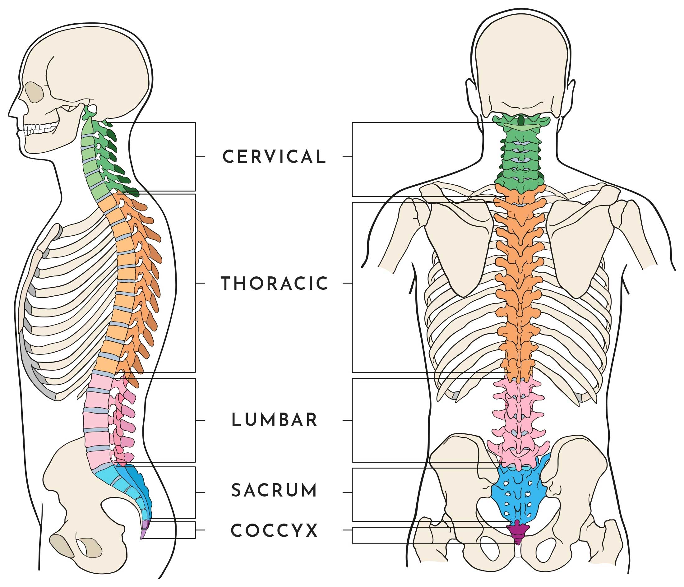
What are the parts of the spine? The spine is made up of 6 key elements, each of which contributes to the function and support that it provides. These elements include: Vertebrae: The bones of the spine. Each vertebra has space in the center, forming a hollow tube when stacked on top of each other so that they protect the spinal canal.
Backbone Drawing at GetDrawings Free download
ISSN 2534-5079. This human anatomy module is composed of diagrams, illustrations and 3D views of the back, cervical, thoracic and lumbar spinal areas as well as the various vertebrae. It contains the osteology, arthrology and myology of the spine and back. It is particularly interesting for physiotherapists, osteopaths, rheumatologists.
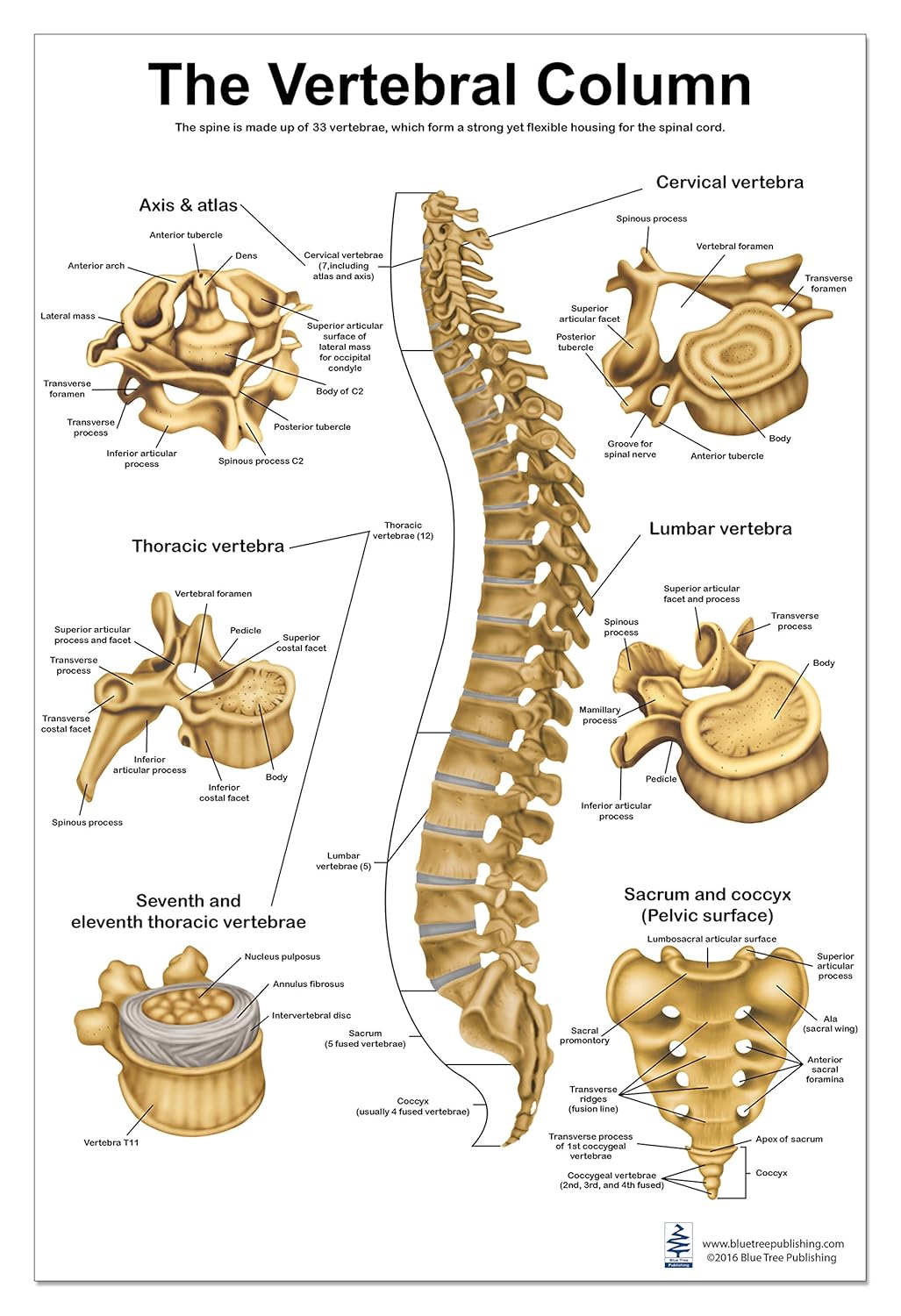
Diagram Of Vertebral Column With Labels
It is the most important structure for any vertebrate. Anatomically, the spinal cord is made up of nervous tissue and is integrated into the spinal column of the backbone. Main Article: Spinal Cord - Anatomy, Structure, Function, and Spinal Cord Nerves Also Read: Central Nervous System - Overview, Parts, and its Functions

Spine Health Tips JOI Jacksonville Orthopaedic Institute
Spine Anatomy Overview Video Typical Anatomical Problems that Cause Back Pain Spinal pain can arise from problems in the bones, nerves, or other soft tissues. Many of the intricate structures in the spine can lead to pain, and pain can be concentrated in the neck or back area, radiate to the extremities, or be referred to other parts of the body.
Labelled Diagram Of Backbone Arthritis of the Neck and the Back Physiatry & HSS Spine A
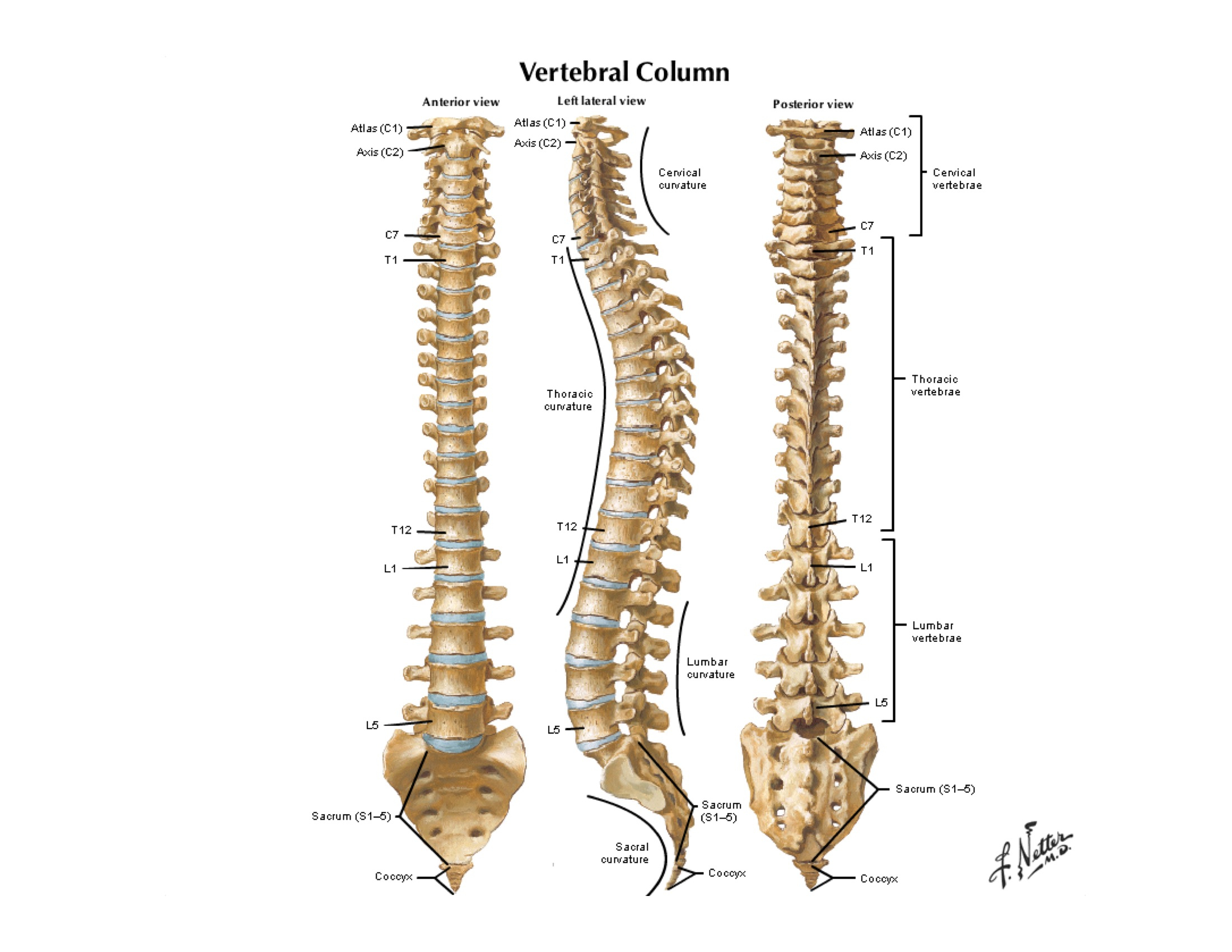
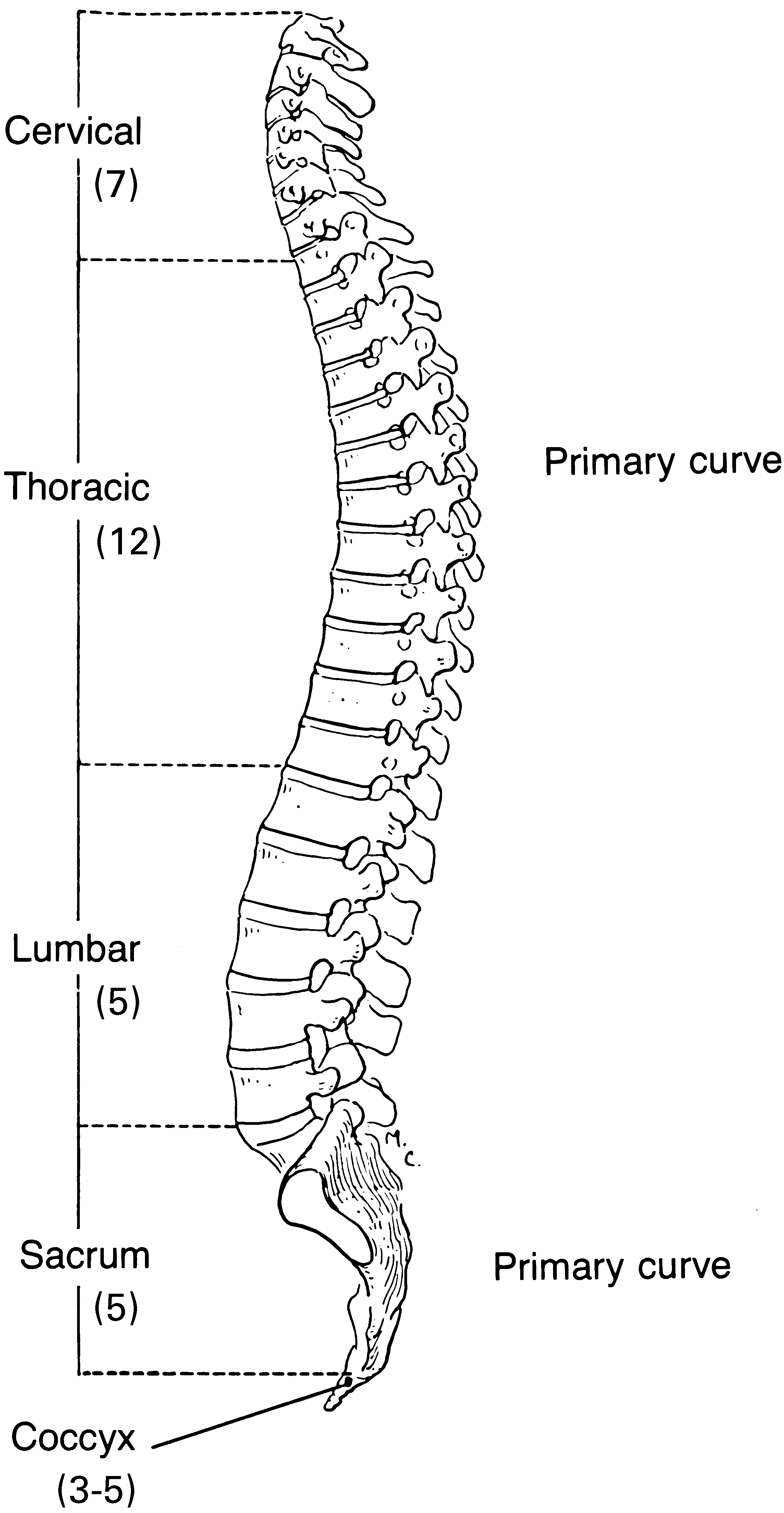
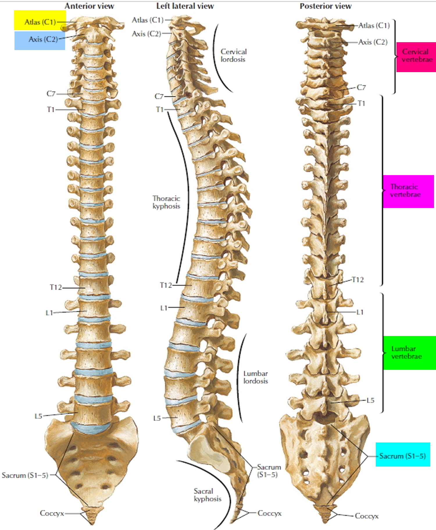
The vertebral column is a series of approximately 33 bones called vertebrae, which are separated by intervertebral discs. The column can be divided into five different regions, with each region characterised by a different vertebral structure.

Diagram of a human spine in front and side Vector Image
The vertebral column, also known as the backbone or spine, is the core part of the axial skeleton in vertebrate animals.. Diagram showing normal curvature of the vertebrae from childhood to teenage. Excessive or abnormal spinal curvature is classed as a spinal disease or dorsopathy and includes the following abnormal curvatures:
Spinal Cord Anatomy Parts and Spinal Cord Functions
What does the lumbar spine do? Your lumbar spine has several functions, including: Supports your upper body, distributes body weight. Your lumbar spine supports the upper two sections of your spine — the seven vertebrae in your neck (cervical spine) and 12 vertebrae in your chest (thoracic spine) — and the weight of your head.

Spine Anatomy Pictures and Information
The gray matter is the butterfly-shaped central part of the spinal cord and is comprised of neuronal cell bodies.It shows anterior, lateral, and posterior horns. White matter surrounds the gray matter and is made of axons. It contains pathways that connect the brain with the rest of the body.. Keep learning about the white and grey matter of the spinal cord using our spinal cord diagram.

Diagram Of Backbone / Anatomy Of The Spine Southern California Orthopedic Institute It keeps
The main bones of the skeleton and their location are shown here: Vertebral column The vertebral column is divided into five main sections and each contains a specific number of vertebrae. There.

Diagram Of Backbone Understanding Spinal Cord Injury Part 1— The Body Before / Backbone
The vertebral column, commonly known as the spine, spinal column, or backbone, is a flexible hollow structure through which the spinal cord runs. It comprises 33 small bones called vertebrae, which remain separated by cartilaginous intervertebral discs. The vertebral column forms the axial skeleton, skull bones, ribs, and sternum.
Anatomy of the Spine
Human body Skeletal System Lumbar Spine Lower Back and Superficial Muscles The muscles of the lower back help stabilize, rotate, flex, and extend the spinal column, which is a bony tower of.
Backbone Drawing at GetDrawings Free download
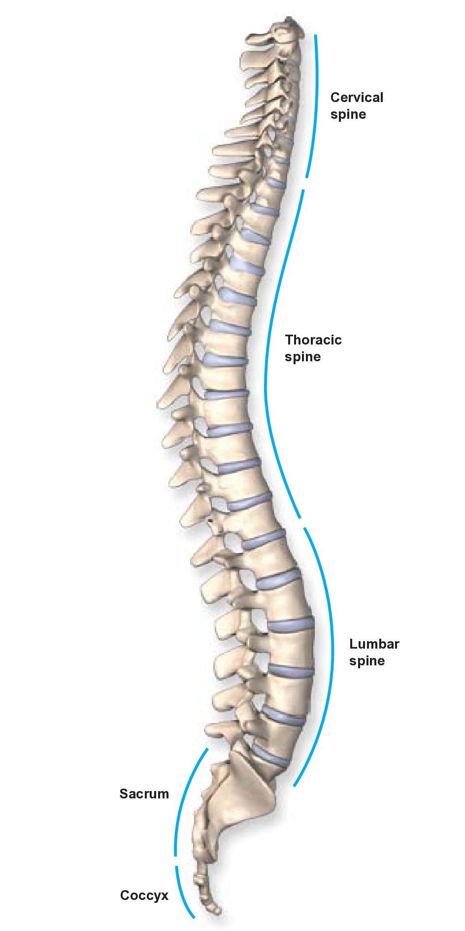
The vertebral column, also known as the spinal column, is a flexible column that encloses the spinal cord and also supports the head. It consists of various groups of vertebrae and is divided.
Anatomy of the Spine Wessex Spinal Surgeon
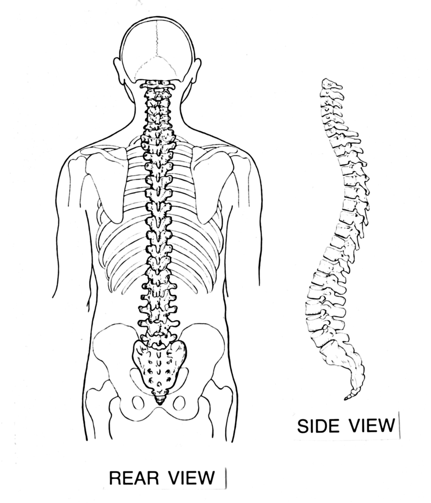
Back anatomy The back is the body region between the neck and the gluteal regions. It comprises the vertebral column (spine) and two compartments of back muscles; extrinsic and intrinsic. The back functions are many, such as to house and protect the spinal cord, hold the body and head upright, and adjust the movements of the upper and lower limbs.

Diagram Of Backbone Antique 1900s Medical Diagram Scientific Print Human / Backbone.js is
Your back consists of a complex array of bones, discs, nerves, joints, and muscles. The muscles of your back support your spine, attach your pelvis and shoulders to your trunk, and provide mobility and stability to your trunk and spine. The anatomy of your back muscles can be complex. There are several different layers of muscles in your back.
